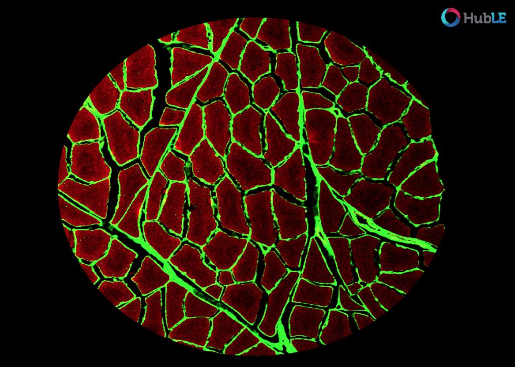
Confocal fluorescent microscope showing tissue cross section of healthy porcine skeletal muscle stained using phalloidin (red muscle cells) and wheat germ agglutinin (green, extracellular matrix).
Image acquired on a Nikon Eclipse E800 microscope with Nikon COOLPIX P600