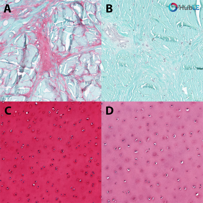
Safranin-O staining of glycosaminoglycan (pink/red) in bovine cartilage after chondroitinase ABC treatment. Top: native (A), and chondroitinase ABC-treated (B) meniscal cartilage; Bottom: native (C), and chondroitinase ABC-treated (D) articular cartilage.

Manula S. B. Rathnayake
The University of Melbourne, Australia
Kathryn S. Stok
The University of Melbourne, Australia


