Early changes in cartilage pericellular matrix micromechanobiology portend the onset of post-traumatic osteoarthritis

Abstract

The pericellular matrix (PCM) of cartilage is a structurally distinctive microdomain surrounding each chondrocyte, and is pivotal to cell homeostasis and cell-matrix interactions in healthy tissue. This study queried if the PCM is the initiation point for disease or a casualty of more widespread matrix degen- eration. To address this question, we queried the mechanical properties of the PCM and chondrocyte mechanoresponsivity with the development of post-traumatic osteoarthritis (PTOA). To do so, we inte- grated Kawamoto’s film-assisted cryo-sectioning with immunofluorescence-guided AFM nanomechanical mapping, and quantified the microscale modulus of murine cartilage PCM and further-removed extracel- lular matrix. Using the destabilization of the medial meniscus (DMM) murine model of PTOA, we show that decreases in PCM micromechanics are apparent as early as 3 days after injury, and that this pre- cedes changes in the bulk ECM properties and overt indications of cartilage damage. We also show that, as a consequence of altered PCM properties, calcium mobilization by chondrocytes in response to me- chanical challenge (hypo-osmotic stress) is significantly disrupted. These aberrant changes in chondro- cyte micromechanobiology as a consequence of DMM could be partially blocked by early inhibition of PCM remodeling. Collectively, these results suggest that changes in PCM micromechanobiology are lead- ing indicators of the initiation of PTOA, and that disease originates in the cartilage PCM. This insight will direct the development of early detection methods, as well as small molecule-based therapies that can stop early aberrant remodeling in this critical cartilage microdomain to slow or reverse disease progression.

Article

Full text of Dr D. Chery’s  article is available from HERE

Capitalising on the use of bone-modulating drugs in the treatment of breast cancer metastasis

Infographic

Abstract

Most breast cancer patients have no evidence of metastasis at the time of original diagnosis, yet ~30% of patients experience recurrent disease. Over 90% of cancer deaths occur due to metastasis, and bone is the most common site of breast cancer metastasis. In some clinical trials, adjuvant zoledronic acid (ZA), a bone-targeting agent, increased disease-free survival but responses were nevertheless limited. The reasons for limited responses are unknown and the mechanisms underlying ZA’s protective effect are unclear. Here, using preclinical breast cancer metastasis models, we establish that bone marrow hematopoietic cells harbor tumor suppressive activity in response to ZA. Specifically, ZA renders myeloid/osteoclast progenitor cells (M/OCPs) tumor-suppressive by altering their gene expression profile and lineage potential. Granulocyte-colony stimulating factor (G-CSF) counteracts ZA’s beneficial effects by directing M/OCP differentiation toward osteoclasts, which ablates metastasis suppression. Women enrolled in a clinical trial who had plasma G-CSF levels >23 pg/mL at randomization experienced a significant reduction in disease-free survival with adjuvant ZA. Our study lays a foundation for understanding breast cancer patient responses to ZA and suggests that finding ways to capitalize on M/OCP function and differentiation potential would constitute novel therapeutic approaches to prevent or limit metastatic disease in the bone.

Thesis

The full text of Dr Jessalyn Ubellacker’s thesis is unavailable. 

Imaging of skeletal muscle cells and extracellular matrix

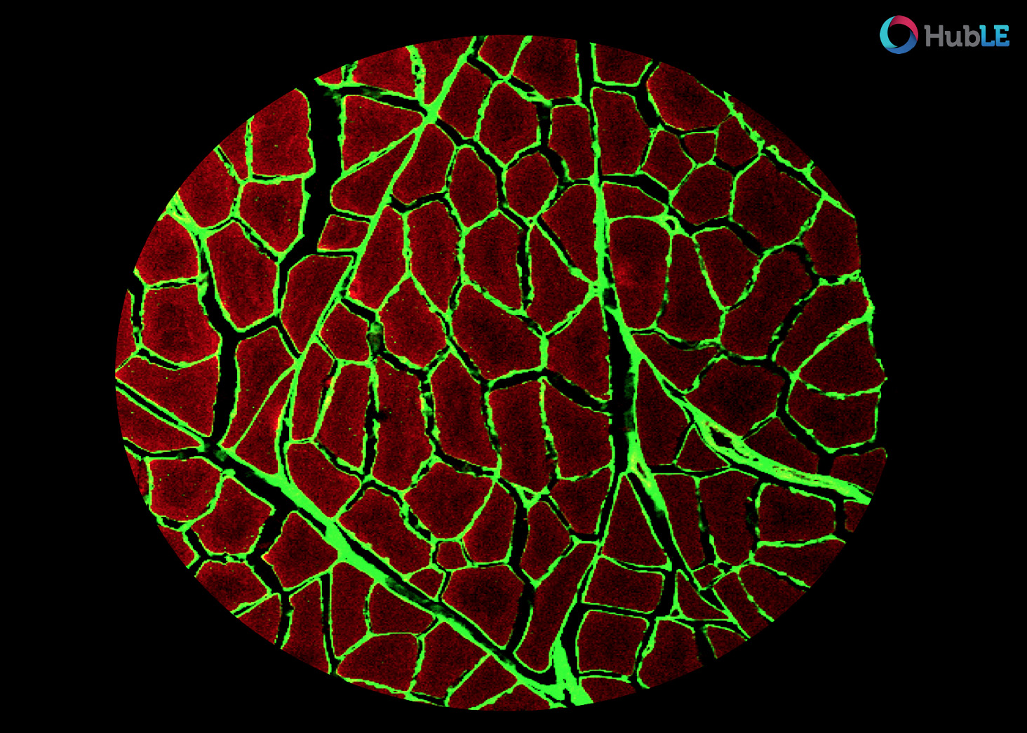
Confocal fluorescent microscope showing tissue cross section of healthy porcine skeletal muscle stained using phalloidin (red muscle cells) and wheat germ agglutinin (green, extracellular matrix).

Image acquired on a Nikon Eclipse E800 microscope with Nikon COOLPIX P600