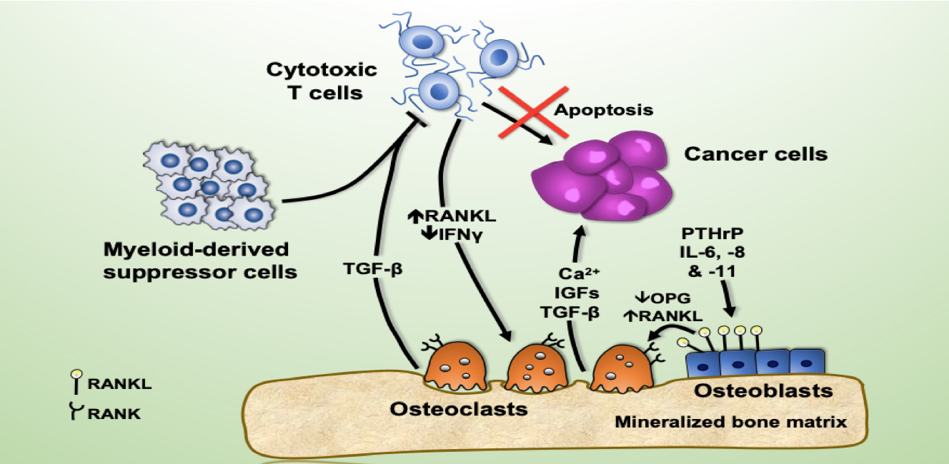
HubLE Graphics

Imaging of skeletal muscle cells and extracellular matrix
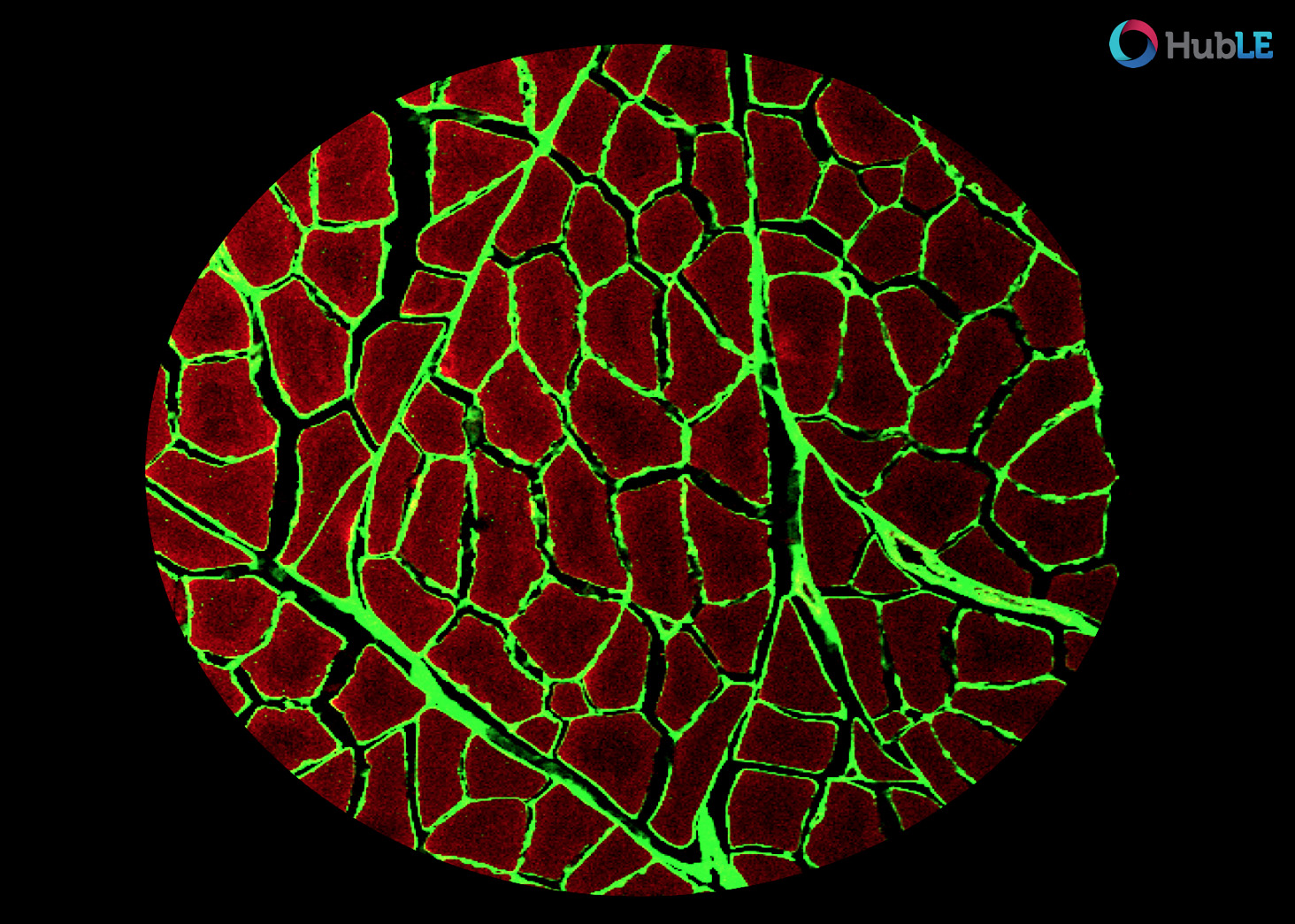
Confocal fluorescent microscope showing tissue cross section of healthy porcine skeletal muscle stained using phalloidin (red muscle cells) and wheat germ agglutinin (green, extracellular matrix).
Image acquired on a Nikon Eclipse E800 microscope with Nikon COOLPIX P600
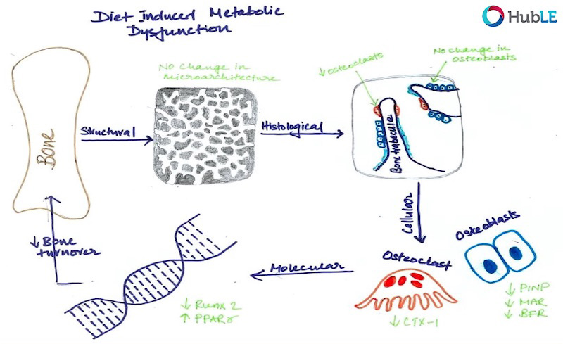
Imaging of prostate cancer dormancy in the skeleton
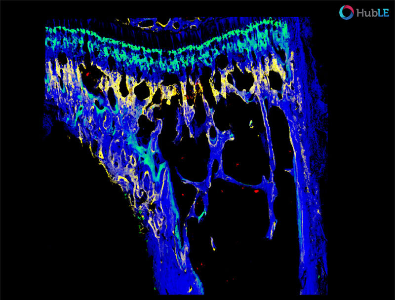
Multi-photo microscopy rendering of prostate cancer cells (red, bone (blue), alizarin complexion (yellow) and calcium (green) at mouse distal tibia.